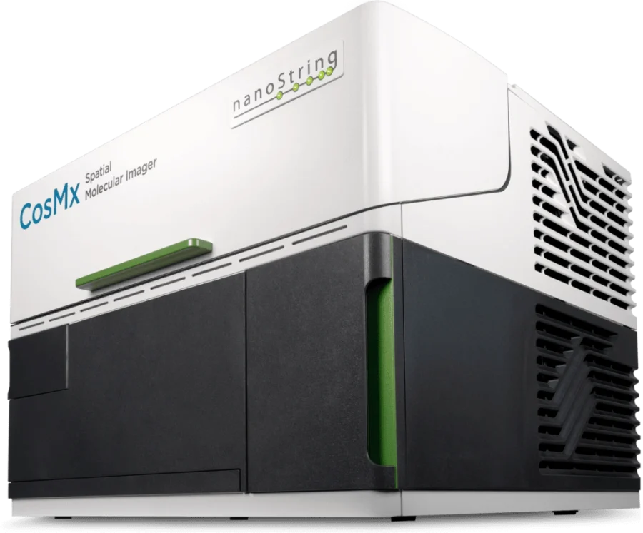
Bruker’s CosMX™ Spatial Molecular Imager (SMI) is an advanced platform for in situ multi-omic spatial analysis at the single-cell level.
CosMx SMI is the only in situ imaging platform that offers single-cell spatial biology of the whole transcriptome with unparalleled accuracy, sensitivity, and genomic breadth. It provides spatial multi-omics on formalin-fixed paraffin-embedded (FFPE) and fresh frozen (FF) tissue samples with single-cell and subcellular resolution.
CosMx SMI enables rapid quantification and visualization of the whole transcriptome (WTX) and 64 validated proteins, offering best-in-class performance for the discovery of unknown and unexpected biology. Capture the history of every cell-to-cell interaction in a whole tissue section with this industry-leading single-cell spatial imaging platform and gain deeper insights for cell atlasing, tissue phenotyping, cell-cell interactions, cellular processes, and biomarker discovery.
Key Features
How does it work?
CosMx SMI is an integrated system with mature cyclic fluorescent in situ hybridization (FISH) chemistry, high-resolution imaging, and interactive software for data analysis and visualization.
 9")
Simple sample preparation, compatible with any sample type
Streamlined and simple workflow that integrates with standard ISH protocols, without the need for tissue expansion or clearing, cDNA synthesis, or amplification. Go from sample to result faster.
 10")
Automated cyclic in situ hybridization chemistry
Robust hybridization chemistry that delivers higher sensitivity and supports high-plex assays on your tissue samples to uncover deeper biological insights.
 11")
Applications
Available Panels
Flexible options to meet your research needs. The major panels of this technology are:
 12")
To learn more about these or other panels, click on the corresponding icon:
 13")
 14")
 15")
 16")