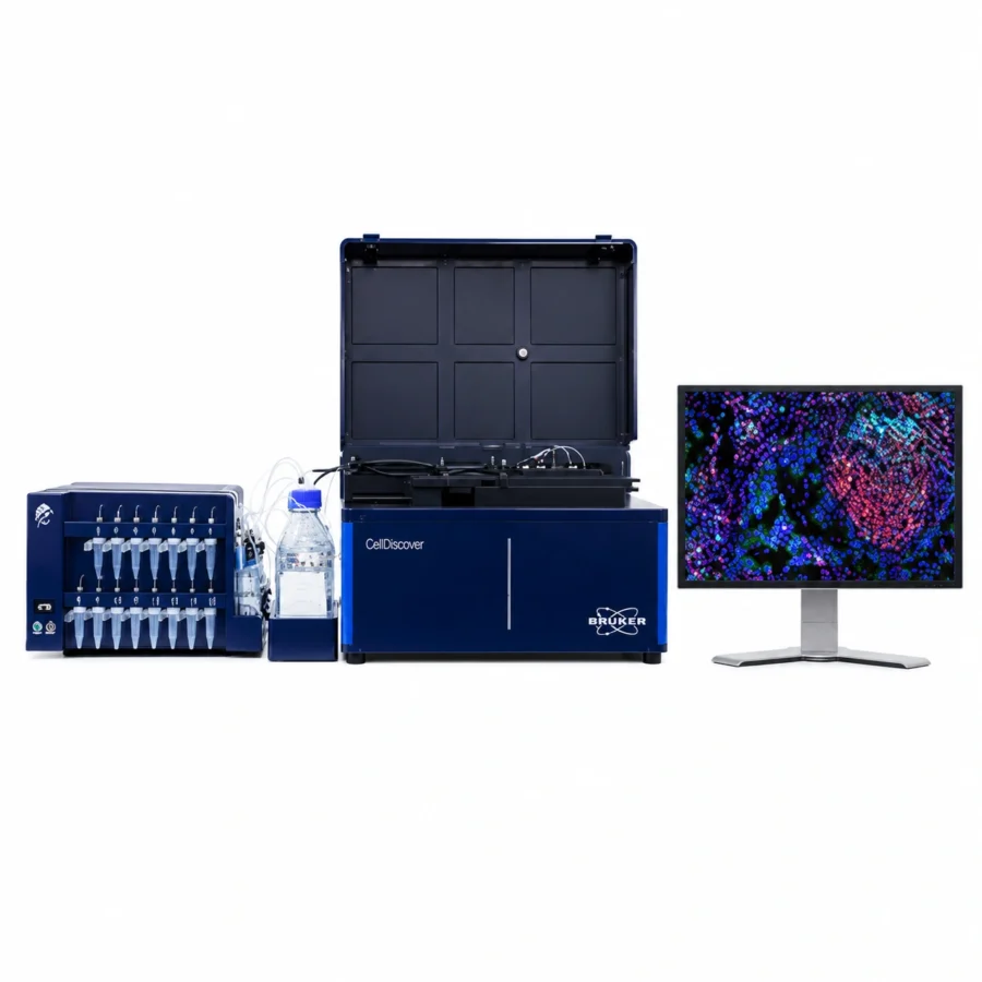
The CellScape™ Precise Spatial Proteomics platform is an optimized multiplexed immunofluorescence (mIF) solution for high-plex spatial protein detection and quantitative phenotyping. By combining an advanced imaging system with integrated microfluidics for “walk-away” automation, and an open ecosystem based on simple chemistry and robust throughput, the CellScape platform accelerates biological discovery and translational research across multiple disciplines.
Key Features
High-resolution imaging with 182 nm/pixel digital sampling.
Spatial proteomics at single-cell and subcellular levels.
High dynamic range (HDR) imaging for accurate detection of low- and high-expressing biomarkers.
Whole-slide imaging across tissue areas >700 mm².
“Walk-away” automated workflow with integrated microfluidics.
Open assay design using commercially available fluorophore-conjugated primary antibodies.
Compatibility with over 6,000 validated antibodies.
Scalable assays through iterative staining and imaging cycles.
EpicIF™ signal-removal chemistry to gently erase fluorescence while preserving tissue.
Standard OME-TIFF outputs compatible with common image analysis software.
Compatible with FFPE tissues, fresh frozen tissues, and cell suspensions.
How does it work?
CellScape™ uses an iterative workflow based on cyclic immunofluorescence. Samples are stained with directly conjugated fluorescent antibodies, high-resolution images are acquired, and then subjected to EpicIF™ signal-removal chemistry to erase fluorescent signals without damaging tissue morphology or epitopes. The process can be repeated multiple times to generate highly multiplexed spatial proteomics datasets from the same sample.
The platform integrates:
Automated microfluidics for reagent dispensing.HDR optical imaging for quantitative biomarker detection.Automatic image alignment between cycles.Flexible assay expansion with additional biomarkers at any stage of the experiment.
This workflow enables precise quantitative phenotyping and spatial analysis of complex tissues at single-cell resolution.
Simple and straightforward workflow from sample preparation to image generation:
Applications
Spatial profiling of tumor microenvironments.Immuno-oncology research.Biomarker discovery and validation.Translational and clinical research.Immune cell phenotyping.Neuroscience research.Infectious disease studies.Cell-cell interaction analysis.Longitudinal and hypothesis-driven studies in spatial biology.Multi-omics workflows integrating transcriptomics and proteomics.
Available Panels: VistaPlex™ Assay Kits
Multiplexed panels of pre-validated antibodies designed for rapid implementation and streamlined assay development.
CellScape™ Data Explorer
CellScape™ Data Explorer is an interactive browser-based platform that allows you to explore real high-plex spatial proteomics datasets generated with CellScape™ and VistaPlex™ Assay kits.
The Data Explorer enables:
Interactive visualization of spatial proteomics datasets directly in the browser.Smooth zooming from full tissue architecture to single-cell details.Individual marker visualization and overlap analysis.Exploration of tissue regions at multiple spatial scales.Phenotype overlay and segmentation for cellular organization analysis.Evaluation of staining specificity and marker localization.Assessment of spatial relationships within tissues.
The representative datasets demonstrate the platform’s ability to combine:
Best-in-class spatial resolution.High dynamic range imaging.Quantitative biomarker analysis.Broad tissue coverage with cell-level detail.
CellScape™ Data Explorer provides a hands-on preview of the depth, quality, and biological insights that the CellScape™ platform can generate across multiple sample types and research applications.